A new high-resolution laser system is being commissioned for performing spectroscopy at the Artemis Analytical facility. This will allow us to more easily differentiate between signals from different isotopes, facilitating the measurement of trace isotope concentrations within a sample.
At Artemis Analytical, our high-speed detection system is capable of measuring single atoms at a time. The challenge lies in making sure we can differentiate signals from atoms of different elements and isotopes. To achieve this, atoms of a particular element are selectively excited and ionised.
Atoms are excited through absorbing electromagnetic (EM) radiation of particular frequencies which are unique to each individual element. EM radiation comes in many forms, from radio waves to visible light, to ionizing x-rays and gamma rays. The type of radiation is defined by its frequency of oscillation.
Lasers are narrow and intense beams of EM radiation traveling in the same direction and oscillating in a very narrow range of frequencies. Higher-resolution lasers have purer frequency content than their low-resolution counterparts – the higher resolution of the laser, the closer it is to oscillating at a single frequency. Although microwave and gamma-ray lasers do exist, most lasers encountered in research and industry are in the visible, infrared and ultraviolet regions of the EM spectrum.
At Artemis Analytical, we overlap beams of fast atoms with a laser beam tuned to the precise frequency to excite atoms of a particular element of interest. When traveling at high speeds, the excitation frequencies of different mass particles are slightly shifted away from each other, meaning that, for example, the excitation frequencies of krypton-84 diverge from those of krypton-85. However, at the acceleration energies used at Artemis Analytical, this shift is small. When measuring an element with one extremely abundant isotope and a neighboring, rare isotope, the peak from the abundant isotope could engulf the peak from the rare one. To distinguish peaks from neighboring isotopes, higher-resolution lasers are preferable. This is because the purer the frequency of the laser, the narrower the measured absorption peak, and the less likely it is that close-together peaks will be indistinguishable.
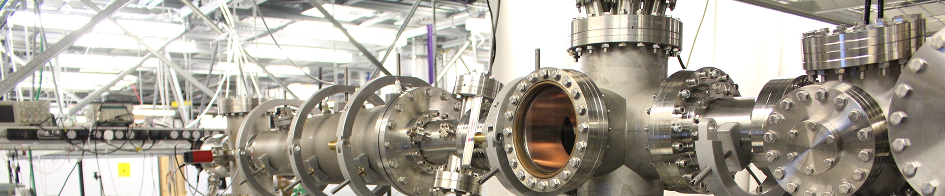
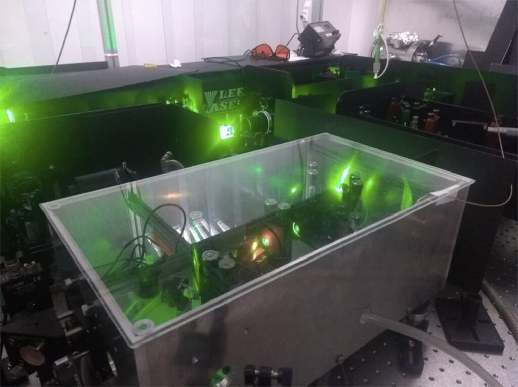
The new laser system uses a titanium sapphire crystal to generate beams in the red and infrared regions of the EM spectrum (a similar system is shown in the photo below). The system requires two auxiliary input laser beams to work. One of these external beams defines the precise frequency of the output. The other sets how many laser pulses per second will be generated, as well as the achievable output power. In the first experiments using this laser, it will be producing 10000 pulses per second (1000 times more pulses per second than the previously used laser system) which will greatly improve our overall efficiency. The beam will be frequency tripled to produce ultraviolet light, with the outlook of repeating our first set of measurements on atomic argon. With the new laser system, we will have the improved resolution and efficiency required to measure the concentration of the rarer argon isotopes, relative to argon-40.
After months of optimisation and development work, the first set of measurements has been made using the Artemis quantum mass spectrometer. This is a huge milestone, as it demonstrates that the components of the instrument are working together as they should be. These preliminary results inform us of what needs to come next in order for the instrument to be employed in industry, measuring the isotopic ratios of radioactive elements in environmental samples.
In ongoing proof-of-principle studies, we are measuring high-lying energy levels of argon atoms, as argon is readily-available and easy to work with. By measuring these energy levels and obtaining values which match those documented in scientific literature, we can verify the functionality of our spectroscopy system. The results also illustrate which areas of the system would benefit from further development. In these studies, our experimental method is continually refined, with the outlook of making the same measurements for a wide variety of elements, and ultimately for a variety of isotopes.
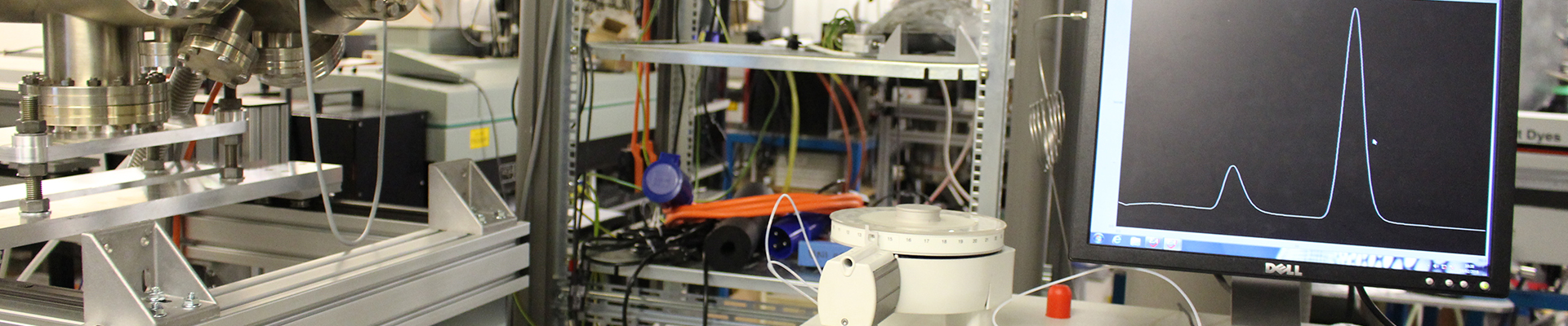
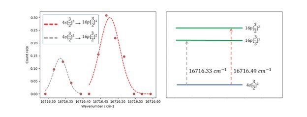
Figure 1: On the left, shows an example of some CRIS data recently collected using the Artemis beamline. The figure on the right demonstrates how the peaks relate to transitions between atomic energy levels. Two peaks in atomic argon are shown, from the metastable 4s[3/2]2 state (to which about 50% of ions are neutralised as they pass through the charge exchange cell) to 2 fine structure components of the n = 16 Rydberg state (given in the key at the top left.) Gaussian profiles have been fit to each peak, the centres of which are 0.01 cm-1 or less away from values of these transitions documented in the scientific literature (Pellarin et al, 1988.) The units of inverse centimetres are a unit of energy, commonly used by spectroscopists.
How it works: the experimental method
In our initial experiments, argon gas is injected into the apparatus and ionised by bombarding the gas particles with high-speed electrons, which boil off a lightbulb-like tungsten filament. The argon ions are accelerated to high energy and guided by electric fields through a cell filled with vaporised sodium metal, which neutralises the incoming ions, producing a beam of argon atoms travelling at high speed (this process is discussed in detail in the previous articles, ‘Fast Atoms‘.)
To measure atomic energy levels, the argon atoms are overlapped with a tunable ultraviolet laser beam. The energy E of a photon is related to its frequency F through the equation E = hf, where h is a fundamental constant of nature. This means that when the frequency of a laser is tuned, the energy of the laser photons is changed too. When the energy of the photons matches the energy required to excite argon atoms to a higher energy level, the atoms can absorb a photon from the laser beam. In atomic physics and physical chemistry, this process is known as resonant excitation. The excited atoms are ionised using another energy source, such as an electric field or another high-power laser beam. The resulting ions are then guided to a detector, where they are counted with a high-speed data acquisition system that is capable of detecting single atoms at a time.
Atoms of each element have a distinct set of quantised energy levels in which their electrons can reside. Electrons usually exist in the lowest energy level available to them, in which case the atom is referred to as being in its ‘ground state’. When an electron is promoted to a higher energy level through absorbing a photon of electromagnetic radiation, the atom is ‘excited’. This can happen if the photon has an energy equal to the energy difference between the upper and lower levels, which is unique to each element, so a photon which excites an argon atom, for example, might not excite an oxygen atom. Atomic energy levels serve as an elemental fingerprint and form the basis for optical spectroscopy.
When an atom is in an excited state, its outer electron resides, on average, further away from the nucleus than it does in a lower state of energy. Because of this, the excited electron experiences less binding attraction to the nucleus and is therefore easier to remove from the atom; in other words, excited atoms are more easily ionised than their lower-energy counterparts. Atoms can exist in a great many different excited states, depending on the amount of energy that is absorbed. In general, the more energy that is absorbed, the further away from the nucleus the electron will reside, and the easier that electron will be to remove from the atom.
Very high-lying energy levels are known as Rydberg states. Rydberg states are a topic of great interest to many chemists and physicists, with relevance in a wide range of fields, ranging from astrophysics to quantum computing. As Rydberg atoms are so easily ionised, this can be achieved using relatively little energy, such as with an electric field produced through applying voltages to a set of electrodes. For us at Artemis, this is a useful tool for element selective analysis. Using a lower energy source minimises the risk that interfering species present within the apparatus will be ionised and measured too, which could otherwise drown out the signals we are looking for.
Field ionisation
In the first phase of experiments, the ionisation step has been achieved by applying an electric field to the region of space through which the excited atoms travel. The field is set up so that the electric field strength changes drastically over just a few millimetres, by applying voltages of between -3000 V and + 3000 V to three extremely delicate wire grids. The grids are composed of carefully wrapped wire which is 10 micrometres thick – less than half the width of a human hair. The more drastically the electric field strength changes over a region of space, the more likely the field is to free an electron from an excited atom.
An imperfect analogy could be tying a balloon (the electron) to the wing mirror of a moving car (the atom). If the car was travelling along at a steady speed and slowed down gradually, the balloon could remain tied to the wing mirror. However, if the brakes were slammed on and the car came to a sudden halt, the balloon would be more likely to break free. The liberating forces originate from the interaction between the electric field and the electrical charge on the electron and nucleus. However, field ionisation is in reality a quantum mechanical process, and the electron acquires enough energy to break free of the atom through quantum tunnelling. The potential energy barrier, which in the absence of an electric field prevents the electron from leaving the atom, is substantially lowered in the direction towards the field anode, so the electron can leave the atom in this direction.
To find the optimal voltages required to ionise the atoms of interest and guide them to detection, simulations of the apparatus are performed. The results of the simulations, an example of which is shown in figure 2, provide approximate values which can be used as a starting point, from which we search for a hint of a resonant ion signal. Once a signal has been detected, the voltages are fine-tuned to maximise the number of ions being measured.
First results
Several transitions in atomic argon have been measured to be consistent with documented values, showing that our apparatus is functioning as required for elemental analysis. From our preliminary spectra, one of which is shown in figure 2, the advantages of field ionisation show up immediately, as mostly 0 ion counts per second are measured in between spectral peaks. This is fantastic news for the future of the apparatus, as it means that when we come to measure very rare isotopes, the small signals from the species of interest will not be drowned out by interfering background ions.
Figure 2: A 3-dimensional plot showing some CRIS data collected at the Artemis beamline, scanning the laser around frequencies corresponding to transitions from the metastable 4s[3/2]2 state in atomic argon to the n=16 Rydberg level. The laser frequency (plotted as ‘wavenumber’) is shown against the number of measured ions per second (on the vertical axis) and the time in microseconds between the moment a laser pulse is fired and the moment the ion is detected. The variety of peaks, each detected at different times relative to the laser pulse, indicates the production of ions via different processes (field ionisation and photoionisation), as well as the presence of species other than argon in the beam. The addition of an electromagnet to the setup will minimize signals from these other species.
From energy levels to isotope ratios
If an environmental sample, such as some krypton gas separated out from the atmosphere, is ionised and formed into a beam, the more abundant isotopes will contribute a greater fraction of the total particles in the beam. When overlapping the neutralised beam with a laser, more atoms of the abundant isotopes will be excited, ionised and subsequently detected than of the rarer ones. This means that, as long as the signals due to the different isotopes can be sufficiently told apart, the relative intensity of these signals (i.e., the relative sizes of peaks arising from different isotopes in a spectrum) can be used to determine the isotope ratios of elements in the sample.
The peaks from different isotopes are already separated due to the fact that the ions are accelerated. Different mass particles travelling with the same kinetic energy travel at different speeds. This means that the laser frequency is doppler shifted by different amounts for each of the different isotopes so that in the lab frame of reference, the excitation frequency of each isotope will be shifted relative to the others. Additionally, in order to rid our spectra of peaks from interfering masses, over the coming weeks a large electromagnet will be installed, which will physically separate the paths of different mass components in the beam.
All in all, it has been an exciting year so far for the scientific team at Artemis analytical, and we greatly look forward to reporting on the upcoming progress over the next few months.
In our last article ‘Beam me Up’ we talked about how we have successfully steered a beam of ions around our new instrument. Here we discuss our next step, how we neutralise the ion beam to produce a beam of fast atoms.
The Quantum Mass Spectrometer will offer highly selective separation of atoms with different masses. For example, we will be able to determine the amount of carbon-14 versus the amount of carbon-12 or carbon-13. The extreme selectivity of the new Quantum Mass Spectrometer is made possible by the interaction of lasers and a beam of fast atoms. At 350 000 km/hr these atoms are fast enough to travel from London to New York in less than a minute.
How is a beam of charged ions converted to a beam of uncharged atoms? The magic happens within a part of the instrument called the charge exchange cell (CEC). A beam of cations (atoms missing an electron) pass into the Charge Exchange Cell where they interact with sodium vapour. Electrons from the sodium atom are passed onto cations in the ion beam converting them back to atoms. The interaction with sodium atoms doesn’t slow the beam, the resulting beam of atoms travel at the same speed as the ion beam.
A charge exchange cell has been successfully installed and tested in our experimental set-up. The cell is loaded with sodium metal, which is vaporised when heated to 250 – 300°C. To heat the charge exchange cell, four 300 Watt cartridge heaters are secured to the body of the cell with metal straps. The heaters are connected together in a parallel circuit, through which a current is passed. This causes the temperature of the heaters to increase.
The temperature of the charge exchange cell is monitored using three thermocouples, which are read using a digital multimeter. One thermocouple is placed against the centre of the cell where the alkali metal resides. The other two thermocouples are secured against each of the CEC’s stainless steel ends. The temperatures of the ends are monitored to ensure that they remain cool enough that alkali vapour condenses at the CEC’s entrance and exit before it has the chance to escape into the apparatus, which would be difficult to clean up and significantly worsen the vacuum pressure.
After installation, the CEC was heated, and a beam of argon ions was passed through the CEC. It was observed that a fraction of the ions were neutralised, verifying that the CEC is working as planned.
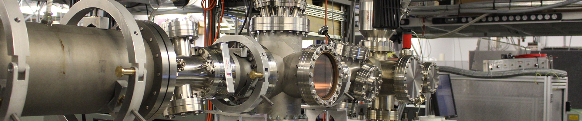
During the initial stages of beamline development, each component is developed, installed and tested individually, to ensure that it behaves as required. During later stages, the beamline components can start to be used together as an instrument. Observing neutralisation of the ion beam is a significant step in the development of our apparatus, as it indicates that the ion source, ion optics, vacuum systems, charge exchange cell and detector systems are all working in concert.
The final pieces of the puzzle are now the laser beams, which will be tuned to manipulate and ionise the atoms of interest and select them from the fast atomic beam. We are currently in the process of setting up the optics necessary to transport the laser beams from their outputs through the instrument, after which spectroscopy experiments can begin.
We’re delighted to announce that we’ve reached a significant milestone on our journey to developing a new trace analysis technique.
Our novel instrument exploits the interaction between a beam of ions that are accelerated through a vacuum and highly tuned lasers. Our team have successfully delivered the first ions to the end of the instrument.
A key step in commissioning a new spectrometer is the process of tuning ions from their source to the end of the instrument. It is always a reassuring moment when ions can be delivered to the final detector and a signal can be observed.
The commissioning process starts with the ion source, which is the beating heart of any mass spectrometer. The performance of the machine, its capability and capacity to deliver science will have a lot resting on having an optimized ion source.
“It has taken some time to get our ion source working optimally and to tune the beamline voltages such that the resulting ion beam will reach the detector. Understanding the relative impact of changing the voltage on each lens and steering element, with the aid of some simple simulations, has been crucial. I was very happy when we finally measured ions on the detector, as this represents an important milestone in the commissioning of this instrument, and is a good indication that all elements in the beamline are working as they should be.” – Holly Anne Perrett, Team Leader
The mass spectrometer Artemis Analytical is developing uses a laser-based method to enhance sensitivity sufficiently to allow rare isotopes such as 14C and 85Kr to be detected. These isotopes have natural abundances of one part per trillion or less, which requires a very intense initial ion beam. Even at moderate ion speeds, there will be enough to make components get very hot (and even melt) if the beam hits them. Therefore, before using intense beams the first step of the commissioning process uses an intermediate intensity ion source, which produces 1/1000th of the final beam current before scaling the system up to the maximum intensities.
We have utilized a device called an electron impact gas ion source that produces beams of argon or krypton for commissioning the instrument. A controlled leak of gas is bled into the ion source to produce almost a trillion ions per second for the commissioning process. The ions are accelerated through a voltage of approximately 1000V and then directed through the vacuum chambers. At these energies, it is possible to use static voltages applied to electrodes.
The process of manipulating the ions is similar to manipulating light with mirrors and lenses and hence the components within a spectrometer are referred to as “ion optics”. To finally deliver the beam to the end of the spectrometer requires adjusting over 25 individual power supplies connected to the ion optic elements to bend and focus the ions.
The first-time ions are accelerated and directed through the instrument is a fiendishly complex puzzle, which is made much harder because unlike manipulating light with optics, there are no visible cues to where the beam is going when it is no longer detected.
This website uses cookies so that we can provide you with the best user experience possible. Cookie information is stored in your browser and performs functions such as recognising you when you return to our website and helping our team to understand which sections of the website you find most interesting and useful.
Strictly Necessary Cookies
Strictly Necessary Cookie should be enabled at all times so that we can save your preferences for cookie settings.
Cookies:
PHPSESSID – Preserves user session state across page requests.
This website uses Google Analytics to collect anonymous information such as the number of visitors to the site, and the most popular pages.
Keeping this cookie enabled helps us to improve our website.
_ga – Registers a uniq e ID th at is used to generate statistical data on how the visitor uses the website
_gat – Used by Google Analytics to throttle request rate
_gid – Registers a uniq e ID th at is used to generate statistical data on how the visitor uses the website
collect – Used to send data to Google Analytics about the visitor’s device and behaviour. Tracks the visitor across devices and marketing channels.
NID – Registers a unique ID that identifies a returning user’s device. The ID is used for targeted ads
If you disable this cookie, we will not be able to save your preferences. This means that every time you visit this website you will need to enable or disable cookies again.